
A 66-year-old male was admitted for Lt hip arthroplasty due to post-traumatic osteoarthritis of Lt hip. He was doing well and denied dyspnea. He had a history of low disease activity psoriatic arthritis and coronary artery disease.
Physical examination:
Chest-xray for pre-operative evaluation was done and it revealed increased interstitial infiltration (as shown above).
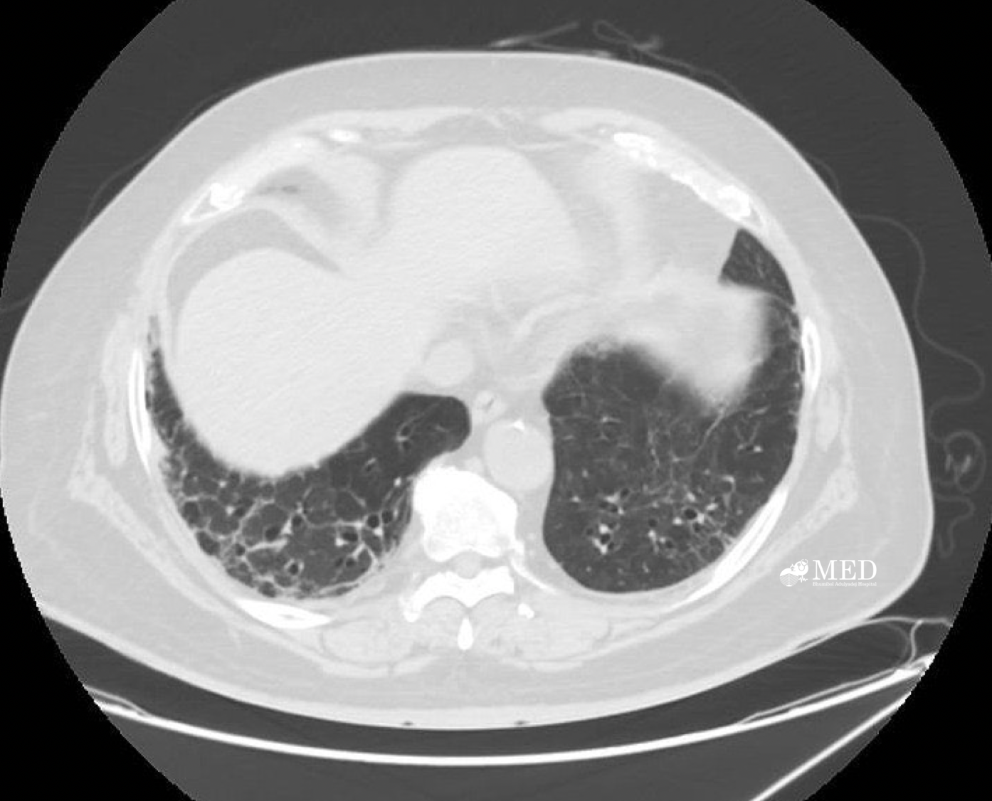
The high-resolution computed tomography (HRCT) scan was not performed due to the inability to schedule it in a timely manner pre-operatively. CT scan of chest was performed (as shown below).

Questions:
1. Describe abnormal findings of CT chest and give provisional diagnosis.
2. Give differential diagnosis of the cause of abnormal CXR and CT chest.
3. What is your plan for next step investigations?
โดย ร.อ.หญิง ณัฐวดี มุ่งการดี แพทย์ประจำบ้านกองอายุรกรรม และ น.อ.หญิง อินทิรา อุไรเลิศ อายุรแพทย์โรคข้อและรูมาติสซั่ม กองอายุรกรรม รพ.ภูมิพลอดุลยเดช กรมแพทย์ทหารอากาศ
ANSWERs